OraGraft® Endure
Oragraft® Endure
OraGraft® Endure
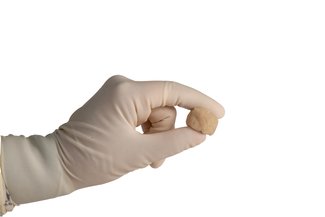
OraGraft Endure is comprised of two components (1) bone fibers which are demineralized to encourage bone formation and healing and (2) cancellous particulate (250-1000 microns) which allows for improved space maintenance. The bone fibers interlock, allowing the graft to become moldable upon rehydration without the use of a carrier.
- 100% Bone: Facilitates natural remodeling during the bone healing process (no human, xenograft or synthetic carriers).
- Osteoconductive: The large surface area and interconnected network of demineralized cortical fibers provides a scaffold that promotes cellular attachment and cell spreading, with the added benefit of space maintenance from the cancellous component.1
- Osteoinductive Potential: Optimally demineralized by LifeNet Health’s patented and proprietary PAD® technology to expose natural growth factors.2-6
- Versatile: Moldable upon rehydration to conform to the surgical site.
- Resists Migration: Interlocking fibers allow graft to remain intact and in place.
- Safety: Sterilized using proprietary and patented technology, providing a sterility assurance level of 10-6 to reduce the risk of disease transmission without compromising the graft’s inherent osteoconductive properties or osteoinductive potential.7
- Convenience: Ambient storage and rapid rehydration.
Clinical Application
Surgical procedures that require a bone void filler
| Freeze-Dried | Description | Sizing |
|---|---|---|
| DFC 1007 | 4 year shelf life | 0.5 cc |
| DFC 1008 | 4 year shelf life | 1.0 cc |
| DFC 1009 | 4 year shelf life | 2.5 cc |
References
- Murphy MB, Suzuki RK, Sand TT, et al. Short term culture of mesenchymal stem cells with commercial osteoconductive carriers provides unique insights into biocompatibility. J Clin. Med. 2013; 2,49-66; doi:10.3390/jcm2030049
- Zhang M, Powers RM, and Wolfinbarger L. Effect(s) of the demineralization process on the osteoinductivity of demineralized bone matrix. J Periodontol. 1997; 68:1085-1092
- Turonis JW, McPherson JC 3rd, Cuenin MF, et al. The effect of residual calcium in decalcified freeze-dried bone allograft in a criticalsized defect in the Rattus norvegicus calvarium. J Oral Implantol. 2006; 32(2):55-62
- Herold RW, Pashley DH, Cuenin MF, et al. The effects of Varying degrees of Allograft Decalcification on Cultured Porcine Osteoclast cells. J Periodontol. 2002 Feb; 73(2):213-9
- Mott DA, Mailhot J, Cuenin MF, et al. Enhancement of osteoblast proliferation in vitro by selective enrichment of demineralized freeze-dried bone allograft with specific growth factors. J Oral Implantol. 2002; 28(2):57-66
- Pietrzak WS, Ali SN, Chitturi D, et al. BMP depletion occurs during prolonged acid demineralization of bone: characterization and implications for graft preparation. Cell Tiss. Bank. 2007 (Published on line)
- Eisenlohr LM. “Allograft Tissue Sterilization Using Allowash XG®.” 2007 Bio-Implants Brief.