
Histological case series of human acellular dermal matrix in superior capsule reconstruction.
Lederman E, McLean, J, Bormann K et al. Journal of Shoulder and Elbow Surgery. 2021
SUMMARY
Acellular dermal matrix (ADM) allografts are commonly used to supplement or reinforce soft tissue repairs, such as rotator cuff. However, limited information is available regarding the biological fate of these grafts in human subjects. This case series describes histological results from eight patients (38-82 y.o.) who had reoperations, during which the previously implanted ADMs were removed, 6 - 38 months after undergoing superior capsule reconstruction (SCR). Each graft’s structure and composition were qualitatively evaluated by one or more of the following histological stains: Hematoxylin & Eosin (H&E), Safranin O, Russell-Movat pentachrome, and Pan-muscle actin.
Grafts showed varying levels of gross and microscopic incorporation with the patient ‘s tissue. An uneven, but high overall degree of recellularization, revascularization, and remodeling was observed. The degree of remodeling correlated with implant duration. The authors conclude that these results represent a significant advancement in our knowledge regarding biological incorporation of ADMs used in SCR and are consistent with successful biological reconstruction of the superior shoulder capsule.
Hospitable scaffold for recellularization:
All ArthroFlex grafts showed some level of recellularization. Cell types included fibroblasts, endothelial cells, and chondrocyte-like cells. No cells associated with inflammation were observed. A strong positive correlation was found between the duration of implantation and percent of graft recellularization [a Pearson correlation coefficient of r = 0.758 (p < 0.05)]. These results show that ArthroFlex acts as a hospitable scaffold for host cells.
Evidence of neovascularization:
Neovascularization, an important aspect of healing, appeared to originate at the periphery of the graft first, and then developed towards the interior. Neovascularization was also observed in newly developed host tissue on the graft surface in some areas.
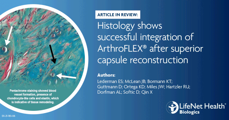
Evidence of remodeling:
Russell-Movat pentachrome and Safranin O staining showed fibrocartilage formation, which is a sign of tissue remodeling. The articular side of the grafts consistently showed high levels of cellularity, whereas the bursal side was inconsistent. Infiltrated cells appeared to have mostly fibroblast-like morphology, with some chondrocyte-like cells mostly found on the articular sides.