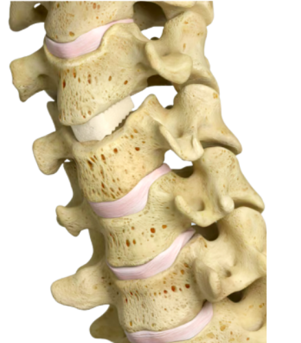
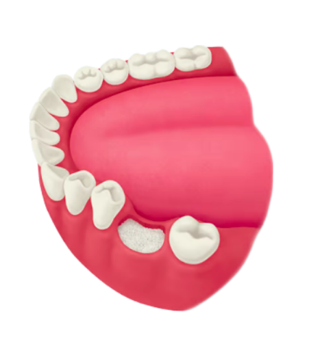
ORIGINAL RESEARCH ARTICLE

OraGraft® Prime – Moldable Demineralized Bone Fibers

Demineralized bone matrices (DBMs) have been used for decades in a wide variety of clinical applications involving bone repair. An ideal DBM provides both osteoinductive and osteoconductive properties, while offering versatile handling capabilities. Additionally, a DBM with angiogenic growth factors further supports new bone formation and healing by facilitating delivery of oxygen and nutrients through new blood vessels. Many commercial DBMs are composed of demineralized bone combined with an inert carrier that is used to improve handling. The proportion of the osteoinductive element of the graft — the demineralized bone — varies widely by manufacturer. In response to this need, LifeNet Health developed OraGraft Prime, an advanced demineralized bone graft that is comprised of 100% bone fibers and offers both osteoinductive and osteoconductive properties, as well as optimized handling. Data from in vitro tests illustrate that OraGraft Prime supports the cellular function and attachment of bone marrow-derived mesenchymal stem cells (BM-MSCs). Scanning electron microscopy (SEM) imaging showed the presence of flattening cells and cell-to-cell interactions after 7 days in culture. Additionally, SEM imaging confirmed the hemostatic nature of OraGraft Prime which is evidenced by the formation of fibrinfibers after mixing with blood. Bone morphogenetic protein (BMP)-2, BMP-7, and osteopontin were found, providing support for the osteoinductive potential of OraGraft Prime. VEGF and angiogenin were also present, indicating OraGraft Prime fibers have angiogenic potential. In vivo data from an athymic mouse model demonstrated the formation of new bone elements and new blood vessels, further indicating the osteoinductive and angiogenic potential of OraGraft Prime. Finally, long-term culturing of fibers with bone-forming cells revealed the graft is readily mineralized as early as 6 weeks. Together, these results suggest that OraGraft Prime provides osteoconductivity, osteoinductive and angiogenic potential, and hemostatic properties to facilitate bone regeneration.

KEY WORDS: demineralized bone matrix; osteoinductive; osteoconductive potential; growth factors; metabolic activity; cell attachment; bone growth


Introduction

The ability of demineralized bone matrix (DBM) to facilitate bone healing has been known in clinical settings for over a century.1 However, it was not until 1965, when Dr. Marshall Urist characterized specific proteins trapped within the bone matrix, that it was understood that bone morphogenetic proteins (BMPs) contributed to the osteoinductive property of DBMs.2 Since the discovery of BMPs, other proteins have also been found to contribute to the process of bone healing and regeneration.3 Angiogenic factors — which initiate angiogenesis, the growth of blood vessels from the existing vasculature — support bone healing by bringing oxygen, nutrients, and bone precursor cells to the injury site.4 DBMs, which use acid demineralization to expose these growth factor proteins, have been used successfully for decades in a variety of complex bone repair procedures.2 In addition to containing active proteins, optimal surface characteristics of the DBM are essential for supporting cellular attachment and proliferation. It is crucial to provide a surface topography that allows for the patient’s own cells to migrate into and proliferate on the scaffold.5-7 These points which support cellular attachment are also ideal for hemostasis, the stopping of blood flow, which is important for controlling bleeding at an injury site.8-10 This is vital as uncontrolled bleeding can result in the necessity of blood transfusion, longer hospital lengths of stay, and increased risk of complications such as infection or reoperations.11,12 

LifeNet Health has developed OraGraft Prime, which consists of 100% bone fibers. These long cortical fibers provide a rough surface with multiple protrusions allowing many points for cellular attachment. The contiguous surface of interconnected fibers allows the cells to spread and make cell-to cell connections. OraGraft Prime is demineralized using LifeNet Health’s patented PAD® technology. Literature suggests that DBMs with different degrees of residual calcium show significant differences in osteoinductivity.8 The proprietary PAD demineralization process results in an optimized level of residual calcium, allowing proteins trapped in the bone matrix to become available to facilitate mineral deposition.8 These proteins include growth factors, such as BMPs, which facilitate the osteoinductive potential of the DBM.2,9 Many manufacturers add synthetic, xenograft, or allograft carriers to improve the handling capabilities of their DBMs; however, it has been reported that some carriers may inhibit osteoinductive potential.10 The optimized handling characteristics of OraGraft Prime are achieved without the use of a carrier. The length and width of OraGraft Prime fibers are designed to encourage malleability, while microhooks allow surrounding fibers to interlock, thereby maintaining placement in the implant site. OraGraft Prime gives surgeons optimized handling capabilities, undiluted osteoinductive potential, and a hospitable scaffold for cellular attachment. This paper reviews the in vitro and in vivo tests that were used to assess the osteoconductive, osteoinductive, angiogenic potential, and hemostatic properties of OraGraft Prime.

Methodology Overview

1. Fiber Generation:

Human cortical long bones were recovered from 6 donors with research authorization through LifeNet Health’s organ and tissue procurement service. The bones were debrided, and marrow and trabecular bone removed. The resulting tissue was processed into demineralized fibers using proprietary procedures developed by LifeNet Health. The demineralized fibers were freeze-dried and sterilized via low-dose, low-temperature gamma irradiation.13,14

2. Fiber Analysis:

a. In vitro Metabolic Activity of Seeded BM-MSCs

Bone marrow-derived mesenchymal stem cells (BMMSCs) seeded on OraGraft Prime fibers were measured for metabolic activity using an alamarBlue™ assay (BioRad, Hercules, CA) over the course of 7 days. BM-MSCs without fibers were also measured and served as the control. This assay was used to determine whether OraGraft Prime fibers served as a hospitable environment to support cellular functions. OraGraft Prime samples from 6 donors were placed in triplicate in low-attachment 24-well cell culture plates and seeded with BM-MSCs. BM-MSCs were seeded at 62,500 cells per 25 ± 1 mg of fiber sample on Day 0 and cultured over 7 days with the appropriate growth media. At 1, 4 and 7 days in culture, media was aspirated and replaced with 1 mL of 10% alamarBlue reagent. After 2 hours incubation, the alamarBlue solution was collected and analyzed to assess sustained cellular viability and proliferation using a fluorescence plate reader. Fluorescence was recorded using relative fluorescence units (RFUs), and values were averaged for each donor lot and normalized to its time-matched control. A one-way ANOVA in conjunction with a Tukey post-hoc was used for statistical analysis.

b. BM-MSC Attachment and Morphology

Scanning electron microscopy (SEM) was used to qualitatively evaluate the attachment and morphology of cells. BM-MSCs were seeded 100,000 BM-MSCs per 25 ± 1 mg fiber sample and imaged at 0.5 hour, 1 hour, 1 and 7 days in culture. Samples were from 2 donors with 4 seeded replicates and 1 fibers-only control for each sample.9

c. Hemostatic nature of demineralized fibers

SEM was used to qualitatively evaluate the interaction of blood with demineralized fibers. A solution of red blood cells in 0.125% CaCl2 was incubated with demineralized fibers for 20 minutes and then immediately fixed with glutaraldehyde prior to imaging.

d. In vitro Growth Factor Analysis

BMP-2 and BMP-7: Samples of OraGraft Prime from 6 donors were digested with collagenase enzymatic solution and assayed for both growth factors. The resulting solutions were analyzed in triplicate using an enzyme-linked immunosorbent assay (ELISA) from R&D Systems, Minneapolis MN. The measured BMP content was averaged across all 6 donors and results were reported in ng protein/g of demineralized fibers.

VEGF, Osteopontin, and Angiogenin: Samples of OraGraft Prime from 3 donors were digested with collagenase enzymatic solution and assayed for osteoinductive and angiogenic growth factors. The resulting solutions were analyzed in duplicate using the MAGPIX® Protein Multiplexing system (Lumenix, Austin, TX), following the manufacturer’s protocol. Growth factor concentrations were reported in pg protein/mL of protein elution.15

e. In vivo Osteoinductive Potential (OI)

OraGraft Prime was assessed in vivo for osteoinductive potential and new bone formation using an athymic mouse intermuscular pouch model. Fiber samples (0.5cc) from 6 donors with research consent (4 replicates for each sample) were prepared and then implanted in the biceps femoris and superficial gluteal muscle of athymic mice. The implants were recovered 5 weeks post-implantation and fixed, sectioned, and stained with hematoxylin and eosin (H&E) for histological assessment.

f. In vitro Mineralization Study

To assess the ability of OraGraft Prime fibers to mineralize, three-dimensional discs (10mm diameter) were constructed from fully processed OraGraft Prime recovered from two donors, rehydrated with saline overnight, and placed in individual wells of a 24 well plate. Primary human osteoblasts (hObs), and human osteosarcoma cells (Saos-2) were seeded onto their respective constructs at 20,000 cells/cc in either basal media or complete media. After six and ten weeks in culture, fibers were fixed with 10% (v/v) formaldehyde, dyed with Alizarin Red S (Sigma Aldrich, Cat. #: A5533) and evaluated for the presence of calcium which is orange to red in color after staining.15


Results

OraGraft Prime supports attachment of mesenchymal stem cells and sustained cellular activity

Overall, the cellular activity of the BM-MSCs was shown to steadily increase during the course of the 7-day investigation. The results indicated that BM-MSCs seeded on OraGraft Prime fibers showed a significant increase in proliferation between days 4 (51.3 ± 1.2 RFU) and 7 (59.5 ± 1.5 RFU) compared to day 1 (21.3 ± 0.8 RFU) (Figure 1). These data suggest that OraGraft Prime provides a hospitable environment for BM-MSCs. Furthermore, SEM images confirmed BM-MSCs attached within 30 minutes of seeding (Figure 2A). At Day 1, imaging showed flattened cells with multiple adhesion points and cellular extensions as well as secretion of extracellular matrix (ECM), which provides a scaffold on which cells can grow and attach (Figure 2B). Additionally, after 7 days in culture, BM-MSCs infiltrated between fibers and demonstrated cell-to-cell interactions, which allow cells to communicate with each other and are critical to the development and function of tissues (Figure 2C). The ability of cells not only to quickly attach to the matrix but also to maintain a healthy morphology throughout the duration of culture provides evidence of the osteoconductive qualities of OraGraft Prime.

Figure 1:

Figure 1

Proliferation of BM-MSCs attached to OraGraft Prime over 7 days.

The average relative fluorescence units (RFU) values for each set of triplicate test samples were normalized to the average RFU of the corresponding control group (fibers of the respective donor cultured without cells) for all six donors. Asterisks represent statistically significant differences from Day 1 proliferation activity.


Figure 2

Figure 2: Representative SEM images illustrating the morphology of cells attached to OraGraft Prime.

Following culture for 30 minutes (A), 1 day (B) or 7 days (C), the samples were fixed in 2.5% glutaraldehyde and processed for scanning electron microscopy. Images are representative of all samples evaluated and were taken at 3000x magnification (scale bar represents 10 μm). Images were pseudocolored in Photoshop to distinguish the cells (in yellow) from the fibers.

OraGraft Prime is hemostatic

SEM images demonstrated that fibrin-like fibers formed after incubation of blood with demineralized bone fibers (Figure 3). The morphology of the erythrocytes was typical in nature, and an abundance of activated platelets were observed.


Figure 3

Figure 3: Representative images illustrating hemostatic nature of demineralized fibers.

SEM images at 3000x (A) and 10,000x (B) magnification fibrin-like fibers were evident, the morphology of erythrocytes was typical, and activated platelets were observed.

OraGraft Prime contains important growth factors and demonstrates osteoinductive and angiogenic potential

In vitro results indicated the presence of osteoinductive growth factors in OraGraft Prime samples (Table 1). The levels of BMP-2 and -7 were consistent with values reported in the literature.16-18 In our study, the average BMP-7 concentration was 85.78 ± 6.84 and the BMP-2 concentration was 11.24 ± 1.49 ng per gram DBM. In further experiments for detecting osteoinductive growth factors, osteopontin was detected in concentrations of 101.49 ± 36.79 ng protein/mL.

Angiogenic growth factors, VEGF and angiogenin, were detected in OraGraft Prime samples (Table 1) at concentrations of 377.49 ± 196.99 and 285.5 ± 171.58 pg protein/mL, respectively. These results demonstrate that OraGraft Prime retains osteoinductive and angiogenic growth factors. Growth Factor Concentration BMP-2 85.78 ± 6.84 ng/g DBM BMP-7 11.24 ± 1.49 ng/g DBM Osteopontin 101.49 ± 36.71 ng/mL VEGF 377.49 ± 196.99 pg/mL Angiogenin 285.50 ± 171.57 pg/mL


Table 1

Table 1: Growth factor content in OraGraft Prime. Osteoinductive growth factors: Using ELISAs for BMP-2 and BMP-7, the resulting digestion solution was tested for BMP-2 and BMP-7 content in triplicate (mean ± SE). Using MAGPIX® Protein Multiplexing system, the resulting digestion solution was tested for osteopontin in duplicate (mean ± SD). angiogenic growth factors: Using MAGPIX® Protein Multiplexing system, the resulting digestion solution was tested for VEGF and angiogenin in duplicate (mean ± SD).

In the athymic mouse muscle pouch model, histological analysis revealed new bone elements and new blood vessels around and within the implanted scaffold at the time of sacrifice (5 weeks; Figure 4). Panel A shows a set of merged images that illustrate new bone elements present in the explant (4x objective). Panels B and C highlight the presence of new bone elements such as cartilage, chondroblasts/cytes, bone marrow, new blood vessels, and new bone.


Figure 4

Figure 4: H&E staining of explants from athymic nude mouse implant with OraGraft Prime.

A) Merged set of H&E images showing new bone elements present in the entire explant at 35 days postimplantation (4x objective). B) and C) H&E images showing the presence of new bone elements such as cartilage (^), chondroblasts/ cytes (#), bone marrow ($), new blood vessels (&), and new bone (+) at 35 days.

OraGraft Prime fibers can be mineralized by bone-forming cells

The in vitro mineralization assay indicated calcium, pigmented red to orange in color by Alizarin Red S, was successfully deposited onto OraGraft Prime fibers by bone-forming cells (hObs and Saos-2; Figure 5A and 5B) as early as 6 weeks and up to 10 weeks in culture with complete media. Control fibers cultured in basal media were not expected to mineralize and showed some red-staining due to residual calcium present after processing. The discs, which were moldable and malleable before incubation with bone-forming cells became rigid after mineralization.


Figure 5

Figure 5: In vitro mineralization of OraGraft Prime Fibers.

Optical microscopy images (4x magnification) of Alizarin Red staining on OraGraft Prime Fibers after 6 and 10 weeks in culture. Fibers were seeded with A) primary human osteoblasts (hObs) or B) human osteosarcoma (Saos-2) cells at 20,000 cell/cc density in either basal media (left column) or complete media (middle and right column). Fibers were fixed with 10% (v/v) formaldehyde before staining with Alizarin Red S dye. Calcium deposits are pigmented orange to red and indicated by white arrows.

Conclusion

In vitro and in vivo data shows that OraGraft Prime exhibits osteoinductive, osteoconductive, angiogenic, and hemostatic properties. OraGraft Prime facilitates BM-MSC attachment, viability, and proliferation, which indicates OraGraft Prime is biocompatible. Fibrinfibers form after incubation with blood which indicates the activation of the blood-clotting cascade. The presence of BMP-2, BMP-7, and osteopontin, important growth factors for bone formation, suggest that OraGraft Prime contains osteoinductive factors. OraGraft Prime also demonstrates angiogenic potential which is supported by the presence of VEGF and angiogenin, both of which promote new blood vessel formation. In vitro mineralization assays provide evidence that these growth factors can promote mineralization by bone-forming cells given the appropriate microenvironment. In vivo data also confirm the osteoinductive and angiogenic potential of OraGraft Prime, with the explants showing new bone elements including cartilage, bone marrow, and new blood vessels.

The results presented here demonstrate that OraGraft Prime exhibits the osteoinductive potential and osteoconductive properties needed to promote bone formation, as well as angiogenic potential and hemostatic properties.


References

1. Senn N. On the healing of aseptic bone cavities by implantation of antiseptic decalcified bone. Am J Med Sci 1889;98:219–243.

2. Urist MR. Bone: formation by autoinduction. Science. 1965; 150(3698):893-899.

3. Lieberman JR, Daluiski A, Einhorn TA. The role of growth factors in the repair of bone. Biology and clinical applications. J Bone Joint Surg Am. 2002;84-A(6):1032-1044.

4. Hankenson KD, Dishowitz M, Gray C, Schenker M. Angiogenesis in bone regeneration. Injury. 2011. 42(6): 556-561

5. Karageorgiou V, Kaplan D. Porosity of 3D biomaterial scaffolds and osteogenesis. Biomaterials. 2005;26(27):5474-5491.

6. Kuboki Y, Takita H, Kobayashi D, et al. BMP-induced osteogenesis on the surface of hydroxyappetite with geometrically feasible and nonfeasible structures: topology of osteogenesis. J Biomed Mater Res. 1998;39(2):190-199.

7. Rodriguez RU, Kemper N, Breathwaite E, et al. Demineralized bone matrix fibers formable as general and custom 3D printed mold-based implants for promoting bone regeneration. Biofabrication. 2016;8(3):035007.

8. Zhang M, Powers RM, Jr., Wolfinbarger L, Jr. Effect(s) of the demineralization process on the osteoinductivity of demineralized bone matrix. J Periodontol. 1997;68(11):1085-1092.

9. Moore-Smith DOL, Robert; Wilson, Anne., Inventor; LifeNet Health, assignee. Pulsatile acidification wave demineralization process for producing osteoinductive bone; and osteoinductive bone thereby. US 6,534,095. 2003.

10. Lee JH, Baek H-R, Lee KM, et al. The Effect of Poloxamer 407-Based Hydrogel on the Osteoinductivity of Demineralized Bone Matrix. Clinics in Orthopedic Surgery. 2014;6(4):455-461.

11. Vera-Llonch M, Hagiwara M, Oster G. Clinical and economic consequences of bleeding following major orthopedic surgery. Thromb Res. 2006;117(5):569-77.

12. Mirza S, Elawady K, Abbas SK, Gangoo SA, Panesar SS. Hemostasis and Biosurgicals in Trauma and Orthopedic Surgery [Online First], IntechOpen. 2020. DOI: 10.5772/intechopen.92805. Available from: https://www. intechopen.com/online-first/72519

13. LifeNetHealth. ES-17-111-02.

14. LifeNet Health. ES-17-110.

15. LifeNet Health. TR-19-0446

16. Bae H, Zhao L, Zhu D, Kanim LE, Wang JC, Delamarter RB. Variability across ten production lots of a single demineralized bone matrix product. J Bone Joint Surg Am. 2010;92(2):427-435.

17. Murray SS, Brochmann EJ, Harker JO, King E, Lollis RJ, Khaliq SA. A statistical model to allow the phasing out of the animal testing of demineralised bone matrix products. Altern Lab Anim. 2007;35(4):405-409.

18. Pietrzak WS, Woodell-May J, McDonald N. Assay of bone morphogenetic protein-2, -4, and -7 in human demineralized bone matrix. J Craniofac Surg. 2006;17(1):84-90.